Cochlear Implantation via the Transmeatal Approach in an Adolescent with Hunter Syndrome—Type II Mucopolysaccharidosis
Article information
Abstract
Type II mucopolysaccharidosis (MPS II) commonly known as Hunter syndrome, is a rare X-linked lysosomal storage disorder caused by iduronate-2-sulfatase deficiency, which in turn causes otorhinolaryngological manifestations, including sensorineural hearing loss (SNHL). Previously, the median survival age of patients with MPS was approximately 13.4 years. However, in the era of enzyme replacement therapy and other multidisciplinary care modalities, the life expectancy has increased. Herein, we report a rare case of an adolescent with MPS II who underwent SNHL treatment with cochlear implantation (CI). Based on unexpected findings of mastoid emissary veins and overgrowth of the vessels around the temporal bone, CI was performed using the transmeatal approach instead of the conventional transmastoid method, to avoid damage to the vessels. The average hearing threshold after CI was 35 dB and no surgical complications were encountered. Adolescent MPS II may present vessel abnormalities, which can reduce the success rate of surgery. In patients with MPS II with SNHL, CI should be performed under careful monitoring of vessel overgrowth. Moreover, with regard to feasibility of CI in adolescent patients with MPS II with SNHL, surgical techniques such as the transmeatal approach should be selected based on adequate assessment of the case.
Introduction
Mucopolysaccharidosis (MPS) is a rare inherited disorder characterized by degradation of glycosaminoglycans (GAGs). Excessive accumulation of GAGs in the tissue alters the cell function that has various clinical consequences [1]. Type II MPS (MPS II) commonly known as Hunter syndrome, is a rare X-linked lysosomal storage disorder caused by a deficiency of iduronate-2-sulfatase, which causes coarse facies, hepatomegaly, airway and pulmonary dysfunction, skeletal deformities, neurological impairment, heart problems, and adenotonsillar hypertrophy [1]. It mainly affects men and boys, and the prevalence is approximately 1 in 100,000 male live births [2].
The Hunter Outcome Survey (HOS) investigated the natural history of hearing loss in MPS II through long-term observational international study of data acquired from 554 patients with MPS II [3]; the results revealed that only 16% of patients included in the HOS entry had normal hearing, while the remaining patients had mild (24%), moderate (31%), severe (22%), or profound (7%) hearing loss, and among them, 25% had pure sensorineural hearing loss (SNHL). Patients with MPS II have a higher incidence of pure SNHL than patients with other types of MPS. In general, these patients appear normal at birth and show development of otolaryngological abnormality, including hearing loss within 2 to 4 years of age. HOS revealed the progressive course of SNHL and increase of the hearing threshold of approximately 1 dB per year [3].
Enzyme replacement therapy (ERT) is the standard treatment for patients with MPS; idursulfase, the recombinant human iduronate sulfatase, is administered weekly [4]. With regard to other treatments such as hematopoietic stem cell transplantation (HSCT), studies have demonstrated that it was effective in improving hearing function. Da Costa, et al. [5] reported that patients with MPS who underwent HSCT achieved a mean reduction of auditory brainstem response (ABR) threshold of 19 dB. Nevertheless, supportive or symptomatic management is an important aspect of the treatment approach; particularly, in case of hearing loss, otologists can improve the quality of life of both the patients and their families. For conductive hearing loss due to otitis media, which is relatively common in patients with MPS, hearing can be improved through insertion of a ventilation tube. However, severe SNHL can also occur in these patients, which requires more advanced management [6].
Cochlear implantation (CI) is an established treatment method for severe SNHL. Saeed, et al. [7] reported their experience of a case of a 4-year-old patient who underwent CI that was effective without intra- or postprocedural complications, and concluded that CI is a useful therapeutic option in patients with MPS II with SNHL. However, studies to assess the performance of CI in older patients with MPS II are needed.
In conventional CI, canal wall-up mastoidectomy and posterior tympanotomy have been used; in addition, a transmeatal approach to insert the electrode array through the external auditory canal (EAC) has been proposed [8]. This technique involves bypass of the mastoid cavity, which can improve the suprameatal approach [9]. The other advantages of the transmeatal approach are as follows: first, there is no risk of facial nerve injury because mastoidectomy is not performed; second, the method can be performed in cases with a narrow facial recess, high jugular bulb, or inferior orientation of the round window niche; third, the transmeatal approach allows direct access to the electrode insertion site through the open tunnel drilled in the EAC [8]. In our study, we used the transmeatal approach for insertion of the electrodes, since we observed prominent vessel overgrowth at the mastoid cavity in our patient, which interfered with the conventional approach.
A study reported that the median survival age of patients with MPS was approximately 13.4 years [10]; however, after the introduction of ERT and other modalities of multidisciplinary care, the life expectancy has increased. Based on these facts, the number of patients with MPS with progressive SNHL presenting at the otologist’s office is expected to increase, including many of those in the relatively older age group. Herein, we report a rare case of an adolescent with MPS II who underwent CI for severe SNHL.
Case Report
A 13-year-old Korean male with MPS II was referred to Department of Otolaryngology, Ajou University School of Medicine, because of progressive bilateral hearing impairment. At presentation, valvular cardiopathy, bilateral equinocavus deformities, kyphosis, developmental language delay, and bilateral hearing loss were detected. Treatment with regular idursulfase injection since 2009 and bilateral hearing aids for approximately 3 years were noted; however, the patient reported low satisfaction with the hearing aids. Ethical review of the study was waived by the Institutional Review Board of Ajou University Hospital (AJIRB-MED-EXP-18-128).
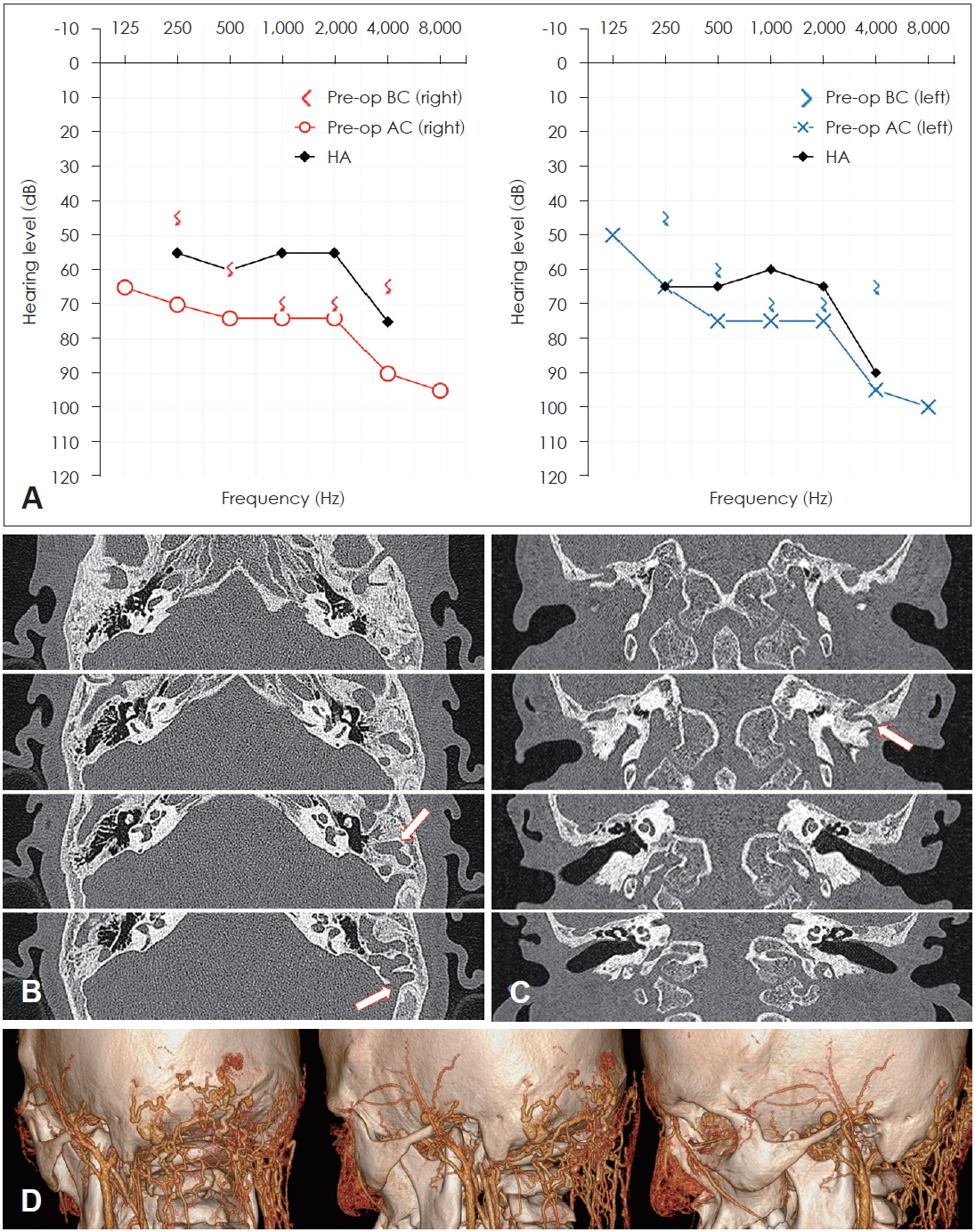
In pure-tone audiometry, hearing thresholds of 75 dB were obtained in both ears (Fig. 1A), and speech discrimination score of 44% at 90 dB was obtained in the right ear and that of 40% at 90 dB was in the left ear. After use of bilateral hearing aids, improvement of those scores of 56% at 65 dB in both ears was observed. In ABR, the hearing thresholds were 70 dB in the right ear and 75 dB in the left ear. In vestibular function evaluation, no specific findings were made.

Preoperative evaluations. (A) Preoperative pure-tone audiometry with or without hearing aids. (B, C) High-resolution temporal bone computed tomography (CT) images at axial and coronal view respectively; white arrows indicate the emissary veins around the left temporal bone. (D) Temporal bone CT with angiographic three-dimensional reconstruction. Diffuse, arterial vessel overgrowth is visualized around the temporal bone. Pre-op: preoperative, BC: bone conduction, AC: air conduction, HA: hearing aid.
On the cochlear magnetic resonance imaging (MRI) and high-resolution temporal bone computed tomography (CT) scans, the presence of normal inner ear structures was observed; moreover, both the mastoid cavities were underdeveloped with poor pneumatization. Suspicious emissary veins were observed in the bilateral portion of the mastoid (Fig. 1B, C). In the oblique sagittal view of MRI, both the cochlear nerves were visualized.
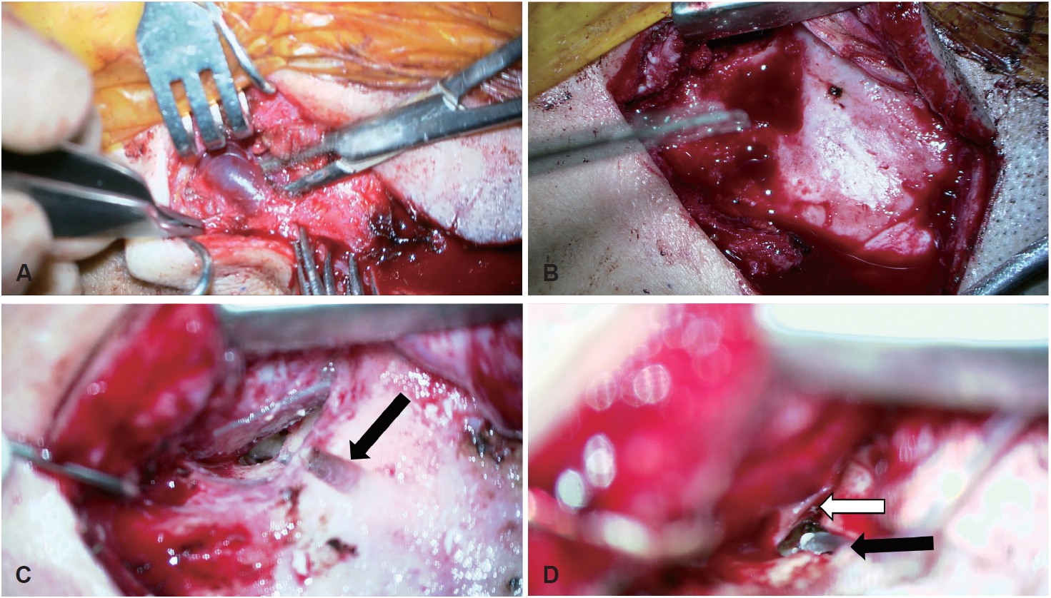
The first operation was performed on the left ear with the conventional transmastoid approach. After posterior auricular skin incision, massive venous bleeding was observed. Substantial vessels in the subcutaneous area were identified during elevation of the skin flap, and the vessels were ligated to access the mastoid bone (Fig. 2A). Careful mastoid drilling was performed using diamond burs, but more vessels were encountered filling the mastoid cavity (Fig. 2B), which could be linked to the sigmoid sinus; hence, a decision was made not to proceed. Finally, the operation was terminated by ligating the vessels and applying bone was to the drilled mastoid bone.

Intraoperative images. (A, B) Intraoperative images of the first operation. Emissary veins are visible between the skin of the scalp and periosteum of the temporal bone. Some vessels derived from the mastoid portion are observed. All vessels around the operation site were ligated or treated with bone wax. (C) Intraoperative images of the second operation. An open tunnel was drilled for the electrode array (black arrow). (D) The electrode array (black arrow) was inserted through the open tunnel, protected by the cartilage (white arrow).
In the preparation stage of the second operation, the following aspects were considered: First, in the results of evaluation of the arterial blood vessels through temporal bone CT with angiographic three-dimensional reconstruction, prominent overgrowth of the arterial vessels was observed (Fig. 1D). Nevertheless, the target surgical area was not compromised by the arterial overgrowth, but the conventional approach with mastoidectomy for insertion of the electrodes was deemed impossible. The transmeatal approach through the EAC was selected to avoid the emissary veins. Second, the implantation site was assessed. Based on the finding that the vessel overgrowth at the mastoid cavity on the right side was relatively less extensive than that on the left side (Fig. 1B, C), implantation in the right ear was initially contemplated. However, we ultimately used the transmeatal approach without mastoid drilling, due to difficulty in the previous surgery caused by the vessels in the subcutaneous area. The overgrowth of emissary veins in the subcutaneous area was difficult to visualize on the CT scan, which prevented clear decision on performing elevation of the skin flap on the right side. On the left side, flap elevation required for the transmeatal approach was partly facilitated by sufficient ligation of the subcutaneous vessels performed in the previous surgery. In addition, considering the future possibility of bilateral CI, decision was made to perform the implantation in the left ear.
In our patient, to avoid drilling for placement of an internal device, screw-type CI device was considered as the best option. Moreover, with regard to the length of the electrode array, length of the array such as 60–70 mm was considered as insufficient to deliver the electrodes from the receiver-stimulator by the transmeatal approach, and the placement site of the receiver-stimulator was obstructed by the presence of emissary veins. Finally, decision was made to use Neuro Zti Implant (Oticon Medical Systems, Vallauris, France), an array of 80–90 mm length.
CI was performed in the left ear 4 months later. Because the vessels in the subcutaneous area were ligated in the previous surgery, no substantial bleeding was observed. After dissection of the skin of the EAC, the posterior EAC wall was partially drilled to form an open tunnel for the electrode array (Fig. 2C). The medial bone segment at the posterior wall of the EAC was also partially removed using curettes and a skeeter drill, which revealed the presence of a pseudomembrane in the region of the round window. The pseudomembrane and round window niche were removed with a skeeter drill, and the electrodes were inserted through the round window. The open tunnel at which the electrode array was positioned was covered with the cartilage (Fig. 2D). Among the total 20 electrodes, 18 were responded to intraoperative neural response telemetry. The electrode array was fixed with fibrin glue (Greenplast-Q kit®, Green Cross, Yongin, Korea) and surgical bone wax. Proper positioning of the electrodes was confirmed in the transocular view (Fig. 3A). No intraoperative or postoperative complications were observed. The patient was discharged without any problems at 2 days postoperatively.

Postoperative evaluations. (A) Postoperative transocular view. (B) Postoperative functional gain test with cochlear implantation. (C) The external auditory canal is patent, and mild anterior protrusion of the cartilage is observed.
At regular follow-up with CI mapping, the average hearing threshold with the implant was 35 dB (Fig. 3B). The surgical site, including the left EAC and tympanic membranes, were fully healed with normal appearance, except for mild protrusion of the cartilage (Fig. 3C).
Discussion
To the best of our knowledge, this is the first report of CI in an adolescent with MPS II. A case of CI in a patient with MPS II was previously reported in the literature, but that patient was only 4 years of age and no anatomical abnormalities were identified [7]. Based on the risks and benefits of treatment using CI, we consider that it is an acceptable option for MPS II with SNHL. However, life-long slow progression of the vessel alterations is a confounding factor of surgical procedures in adolescents with MPS II. The current report highlights the difficulties associated with CI surgery in an adolescent patient with MPS II.
Patients with MPS often exhibit hearing impairment. In childhood, conductive or mixed-type hearing loss caused by otitis media is common, but the relative incidence of pure SNHL increases with age [1,6]. Moreover, the medical issues of patients with MPS are changing due to early diagnosis and increase of life expectancy, multidisciplinary care, and the introduction of ERT and HSCT [5,10,11]. Today, MPS patients live long enough to develop hearing problems. Because of these increases, combined with progressively deteriorating hearing function, more MPS patients with hearing loss require additional management by otologists [3,6].
Studies have indicated that CI is a useful option for MPS patients with SNHL [7]. In our study, the patient was a 13-year-old male who showed atypical vessel overgrowth in the temporal bone area, which was the main contributing factor for difficulty of the operation; in addition, transmastoid emissary and extracranial veins drained to the sigmoid sinus. A study has reported similar vessel abnormality in patients with CHARGE syndrome [12]. Ganaha, et al. [13] described the performance of CI in a patient with CHARGE syndrome with venous anomalies; those authors used a suprameatal approach with cartilage protection. However, there are no reports of such venous malformation in patients with MPS. In general, patients with MPS II show cardiovascular problems [4,10,11]. GAG deposition in the coronary arteries triggers narrowing or occlusion and can even cause sudden death [14]; since GAGs are deposited at various sites in the entire body, patients with MPS may show involvement of other vessels. One study reported the cerebrovascular changes of a patient with MPS IVa [15], but no reports of cerebrovascular vessel overgrowth in patients with MPS II are currently available. We observed obvious collateral arteries, and both the emissary veins and collateral artery overgrowth were attributable to vessel narrowing caused by GAG deposition. Currently, the life expectancy of patients with MPS is longer, which may influence surgical interventions used in the treatment of adolescents or adult patients, and unexpected vessel overgrowth may compromise the success of surgery. Based on our experience, we propose that combined CT and angiographic reconstruction should be performed preoperatively.
Profuse bleeding from the transmastoid emissary veins may occur, as we observed in the first surgery; therefore, we selected the transmastoid approach to the round window as an alternate method to the conventional facial recess approach with mastoidectomy. According to the method of Ganaha, et al. [13] we created in EAC open tunnel for placement of the electrode array; subsequently, drilling of the medial segment of the EAC posterior wall attained exposure of the round window with relative ease in our patient. However, round window exposure may not be readily achieved in all patients, and since it is a main potential factor of the success of CI through the transmeatal approach, surgeons should consider this issue while planning surgical approach. We used the cartilage to protect the electrode array from contact with the skin. A follow-up after surgery, we confirmed that patency of the EAC was maintained and the electrodes’ function was normal. A study including 131 patients who underwent CI using the transmeatal approach with follow-up of 2 to 46 months reported no electrode extrusion in the cartilage area [8]. A study with long-term follow-up is necessary to confirm favorable outcomes at the cartilage site.
The vessel overgrowth around the area of the receiver-stimulator is a challenging issue. To avoid the risk of bleeding, we were unable to drill the well for the receiver-stimulator to the preferred depth. The position of the screw retaining the internal device were chosen to avoid vessel injury. For selection of a CI device with an electrode array of appropriate length for patients with MPS II, surgeons should consider the location of the receiver-stimulator and its distance from the electrode insertion site. For our patient, we considered that the 80 to 90-mm-long array was appropriate, among the arrays of various length provided by different manufacturers.
Adolescent patients with MPS II may present atypical finding, as compared to those younger patients undergoing CI at early age, as in the previous case report. In CI for patients with MPS II with SNHL, surgeons should consider the possibility of vessel overgrowth. Adequate assessment is needed to determine the most appropriate surgical technique, such as the transmeatal approach, which enables the choice of CI as a feasible treatment option in adolescent patients with MPS II with SNHL.
Notes
Conflicts of interest
The authors have no financial conflicts of interest.
Authors’ contribution
Conceptualization: Yun-Hoon Choung. Data curation: Hantai Kim and Jun Young An. Investigation: Hantai Kim, Jun Young An, and Oak-Sung Choo. Supervision: Yun-Hoon Choung. Visualization: Hantai Kim. Writing of the original draft: Hantai Kim and Yun-Hoon Choung. Review and editing of the manuscript: Jeong Hun Jang, Hun Yi Park, and Yun-Hoon Choung.
