


Translabyrinthine Excision of a Transmodiolar Intralabyrinthine Schwannoma Mimicking Meniere’s Disease: A Case Report
-
Sunil Goyal
 , Mahesh Ravunnikutty, Himanshu Swami, Sneha Yadav
- Received July 14, 2023; Revised August 19, 2023; Accepted September 17, 2023;
- ABSTRACT
-
Intralabyrinthine schwannomas (ILSs) are rare tumors involving the otic capsule. Notably, they are often misdiagnosed because their symptoms mimic those of other, more common inner ear pathologies. Diagnosis requires high-resolution contrast-enhanced magnetic resonance imaging (MRI), which reveals filling defects (using a T2-weighted MRI sequence) or focal enhancement (using a T1-weighted MRI sequence with gadolinium enhancement) in the inner ear. A 52-year-old male patient with intractable vertigo or single-sided deafness should raise suspicion of this clinical entity as a differential diagnosis. Translabyrinthine excision of the tumor along with auditory rehabilitation using a cochlear implant can provide good outcomes with minimal morbidity in carefully selected cases. Here, we present an interesting case of a transmodiolar ILS mimicking Meniere’s disease, wherein surgery using the translabyrinthine approach and an extended cochleostomy yielded favorable outcomes.
- Introduction
- Introduction
Vestibular schwannomas are benign neoplasms originating from perineural Schwann cells of the eighth nerve and commonly develop in the internal auditory canal (IAC) and cerebellopontine angle (CPA) [1]. They rarely arise within the inner ear and are then termed intralabyrinthine schwannoma (ILS). They have a unique clinicoradiological presentation and pathology and may be identified in the vestibule or cochlea or both and may extend to IAC and CPA [2] or middle ear. Kennedy, et al. [3] classified them into intravestibular, intracochlear, intravestibulocochlear, transmacular, transmodiolar, translabyrinthine, and tympanolabyrinthine tumors. Van Abel, et al. [2] modified it by including tumors extending to CPA. Patients generally present with unilateral sensorineural hearing loss (SNHL) or mixed hearing loss, tinnitus, and vertigo. They require high-resolution magnetic resonance imaging (MRI) for diagnosis. Usually, patients are followed up with serial MRI. However, surgery is now commonly applied. Here, we present an interesting case of ILS mimicking Meniere’s disease with involvement of vestibule, cochlea, and transmodiolar extension into the IAC which was managed surgically with satisfactory results.
- Case Report
- Case Report
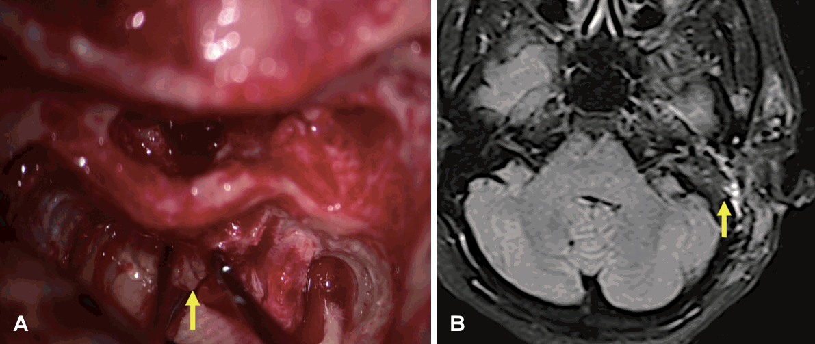
A 52-year-old male patient was referred to our center with complaints of progressive hearing loss and tinnitus in the left ear for the last 6 years and intermittent recurrent rotatory vertigo for the last 10 months. Each episode of vertigo lasted 30 minutes to a few hours and required frequent use of labyrinthine sedatives. The vertigo was not controlled with betahistine or vestibular rehabilitation exercises. The general and systematic examinations were within limits. Otoscopy revealed bilateral intact and mobile tympanic membrane. Tuning fork test was suggestive of profound SNHL left ear. Fistula test and otoneurological examination were normal. Rest of otorhinolaryngologic examination was normal.Pure tone audiometry revealed profound SNHL in the left ear. Impedance audiometry revealed bilateral type A curves and absent ipsilateral acoustic reflex on the left side. In view of asymmetrical SNHL with intractable vertigo, the patient was advised contrast-enhanced MRI (CE-MRI) of brain and inner ear which revealed homogenous enhancing lesion involving cochlear basal turn (5 mm×2 mm), vestibule (3 mm×3 mm), and IAC (5 mm×2 mm) as depicted in Fig. 1.Based on the clinicoradiological findings, he was diagnosed as a possible ILS (transmodiolar type).In view of IAC extension, intractable vertigo, and profound hearing loss, the patient was offered surgical excision of tumor. He underwent excision of tumor via translabyrintine approach with an extended cochleostomy under general anesthesia. Written informed consent was taken. Cortical mastoidectomy was done via postauricular approach. Posterior tympanotomy and cochleostomy were done and tumor involving basal turn was removed and sent for frozen which revealed schwannoma. Next semicircular canals were delineated (Fig. 2) followed by labyrinthectomy. Tumor was seen involving vestibule and was removed. On further tracing, tumor was seen eroding the modiolus (transmodiolar) and extending to IAC. The IAC was delineated from fundus to porus. IAC dura was exposed and excised, and tumor was found to be extending along inferior vestibular nerve (IVN), cochlear nerve (CN), and modiolus (Fig. 3). Tumor along the IVN, CN, modiolus, and basal turn of cochlea was excised preserving the anatomical integrity of facial nerve (Fig. 4). The defect was closed in layers with fascia lata and fat. Fibrin sealant (Baxter Tisseel) and oxidized regenerated cellulose (Ethicon Surgicel) were used additionally for hemostasis.Immediately after surgery, the patient developed House–Brackmann grade 4 facial palsy, which gradually improved to grade 2 with oral steroids over next 2 weeks. Postoperative CE-MRI on day 1 following surgery revealed no residual tumor (Fig. 4). Patient is on regular follow-up for past 2 years and has been free of vertigo with complete recovery of facial nerve function. Follow-up CE-MRI was conducted at 6 months and 1 year, revealing no residue or recurrence.
- DISCUSSION
- DISCUSSION
ILSs are generally slow growing rare tumors of inner ear and pose a low risk of extending to intracranial structures. Symptomatic patients may mimic Meniere’s disease, sudden SNHL, vestibular migraine, and viral labyrinthitis leading to misdiagnosis. Moreover, these subtle lesions can be easily overlooked on MRI [3] of brain and inner ear. The densely located Schwann cells at the glial Schwann junction in the modiolus explain the origin of ILS [2].Clinically, patients may present with vertigo and unilateral hearing loss. Vertiginous symptoms generally manifest in patients when tumor involves vestibule or semicircular canals [3]. The vertigo experienced in ILS (of vestibule or semicircular canals) differs from the unsteadiness felt in patients with schwannomas of the IAC. This distinction likely arises because ILS likely originate from nerve branches within the vestibule, avoiding damage to neural cell bodies in Scarpa’s ganglion in the IAC. Consequently, certain vestibular neurons can still transmit unusual signals to the brainstem and cerebral cortex. The tumor’s presence directly stimulates the vestibular end organs, causing abnormal sensory perception of motion. Also, the slow growth of tumor often allows for central compensation, alleviating vertigo leading to episodic symptoms [4]. Zhang, et al. [5] demonstrated the presence of endolymphatic hydrops in ILS patients, attributed to the mechanical obstruction of endolymphatic flow, resulting in symptoms mimicking Meniere’s disease.Hearing loss is usually unilateral SNHL and can vary in onset from being sudden or gradually progressive or fluctuating leading to clinical dilemma. Mixed hearing loss is seen in intracochlear type extending to middle ear via round window or intravestibular type causing stapes footplate fixation. Audiological evaluation is done to confirm the type and severity of hearing loss. Videonystagmography may show decreased responses on the affected side [2,6]. In the present case report, the patient had clinical presentation of insidious onset gradually progressive unilateral SNHL with tinnitus and episodic rotatory vertigo mimicking Meniere’s disease. The vertigo was intractable as it was not responding to medical management.The diagnosis of these tumors may be delayed or missed as they mimic other inner ear conditions and due to small size of the tumors. High-definition CE-MRI is imaging modality of choice. On constructive interference in steady state T2-weighted MRI, filling defects are seen, and on gadolinium-enhanced T1-weighted MRI, focal enhancement is seen [1]. ILS is identified as a well-marginated focal enhancement on MRI.Management of ILS is determined by the severity of symptoms and location of tumor, which includes wait and scan or surgical excision. Patients with serviceable hearing and controlled vertigo can be observed with serial audiometry and MRI scans. Surgery used to be mainly indicated for intractable vertigo, tumors extending to IAC or middle ear, or serial imaging shows increase in size and uncertainty regarding diagnosis [2,3]. However, in contemporary practice, surgery is employed even for instances of unserviceable hearing loss that do not involve vertigo. Plontke, et al. [7] have reported the maintenance of vestibular function in patients who underwent surgical excision of intracochlear tumors. Objective clinical assessments in the majority of cases revealed proper functioning of the vestibular system despite significant surgical impact on the cochlea [4]. In the present reported case, surgery was indicated because of intractable vertigo with profound hearing loss and tumor extension into the IAC.Patients should be counseled about non-serviceable hearing following surgery. Tumors limited to vestibule and IAC can be removed via transmastoid translabyrinthe approach. While tumors involving cochlea and middle ear require a transotic approach. Tumors minimally involving cochlea are removed via facial recess approach or endaural endoscopic approach [3,6]. In the present reported case, the patient underwent surgery via translabyrinthine approach with extended cochleostomy. Intraoperatively, the tumor was found involving vestibule, basal turn of cochlea with erosion of modiolus, and transmodiolar extension into IAC.Evidence shows that hearing loss including single-sided deafness significantly affects the quality of life and leads to increased risk of dementia and cognitive loss in later life. Hearing rehabilitation with cochlear implantation (CI) is now commonly contemplated in such cases. In studies conducted by Plontke and Collegues [7,8], a trend was noticed wherein patients with sporadic intracochlear and intravestibulocochlear inner ear schwannomas displayed improved hearing outcomes through CI when compared to control groups of other CI patients. This trend persisted even in the presence of significant surgical trauma resulting from tumor removal, and even after long periods of deafness. ILS with transmodiolar extension presents a rare entity. CI becomes feasible only when the tumor removal is partial, as complete excision would entail damaging the spiral ganglion cells within the modiolus [9]. We are a government institution offering cost-free treatments, and CI is not permitted for cases of single-sided deafness. Therefore, complete removal of tumor involving cochlear modiolus and basal turn was done. The patient was given a soft band trial of the bone conduction implant (BCI) and showed satisfactory performance. The patient has given consent for the BCI and is awaiting surgery, subject to the availability of the implant.After surgery, the patient is followed up with CE-MRI to rule out residual disease or recurrence. Follow-up MRI is done generally after 12 months post-surgery as an earlier scan may show enhancement due to postoperative inflammation during healing [10]. Stereotactic radiosurgery is considered an alternative option for elderly patients or those unfit for general anesthesia. However, its application is limited. Radiotherapy also damages neuronal structures leading to cochleovestibular dysfunction and unfavourable prognosis for future CI. Transtympanic gentamicin injections can be considered in patients unwilling for surgery for intractable vertigo [11].Complications are less reported. Facial paralysis reported following removal of tumor with significant IAC extension generally improves with conservative management. Other complications include CSF leak and meningitis [6]. In the case report, the patient had a postoperative facial paresis, despite identifying and preserving the anatomical integrity of facial nerve during surgery, which improved with conservative management.ILSs are often misdiagnosed because of the symptoms mimicking other more common inner ear pathologies. Based on this case, clinicians should consider this clinical entity as a differential diagnosis in patients presenting with intractable vertigo or hearing loss. Transabyrinthine excision of these tumors coupled with hearing rehabilitation using CI can provide good outcomes in carefully selected cases with minimal morbidity.
- Notes
- Notes
-
Ethics Statement This case has been reported with the approval of the Institutional Ethical Committee of Army Hospital Research and Referral, Delhi, India (no. 71/019) and informed consent of the patient.
- Conflict of interest
- Conflict of interest
-
Conflicts of Interest The authors have no financial conflicts of interest.
- Notes
- Notes
-
Author Contributions Conceptualization: Sunil Goyal. Data curation: Mahesh Ravunnikutty, Sneha Yadav. Formal analysis: Sunil Goyal, Himanshu Swami. Investigation: Sunil Goyal. Methodology: Sunil Goyal, Himanshu Swami. Project administration: Mahesh Ravunnikutty, Sunil Goyal. Supervision: Himanshu Swami. Validation: Himanshu Swami. Writing—original draft: Mahesh Ravunnikutty, Sunil Goyal, Sneha Yadav. Writing— review & editing: Mahesh Ravunnikutty, Sunil Goyal, Himanshu Swami. Approval of final manuscript: all authors.
- Notes
- Notes
-
Funding Statement None
- Acknowledgments
- Acknowledgments
None
Fig. 1.
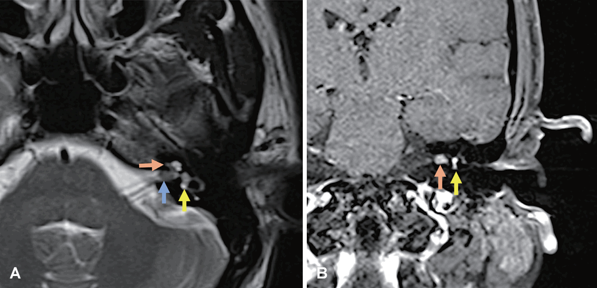
Fig. 2.
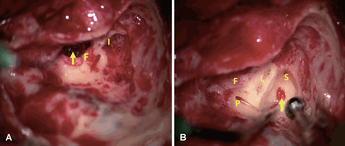
Fig. 3.
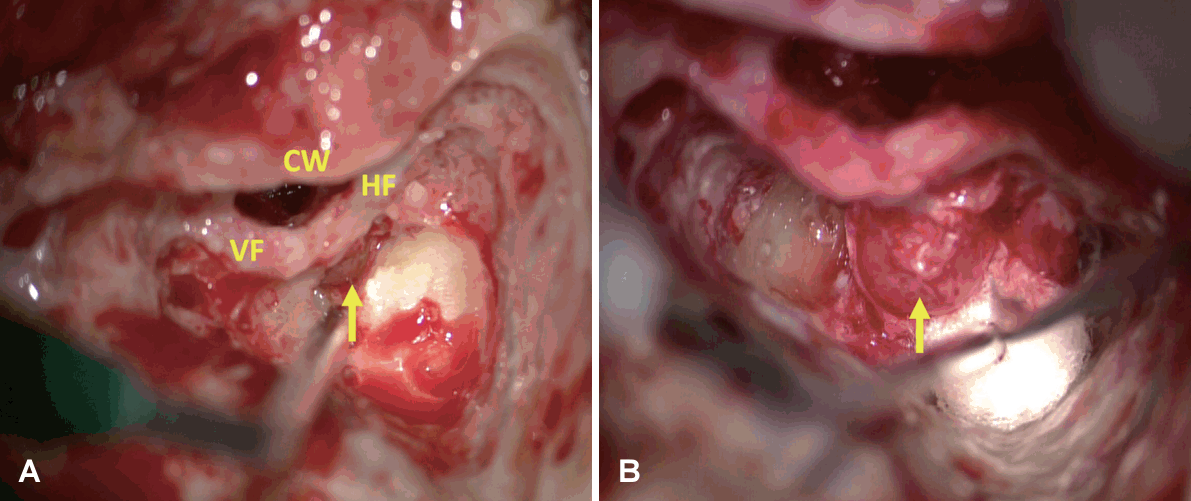
Fig. 4.
- REFERENCES
- REFERENCES
- 1. Salzman KL, Childs AM, Davidson HC, Kennedy RJ, Shelton C, Harnsberger HR. Intralabyrinthine schwannomas: imaging diagnosis and classification. AJNR Am J Neuroradiol 2012;33:104–9.
[Article] [PubMed] [PMC]2. Van Abel KM, Carlson ML, Link MJ, Neff BA, Beatty CW, Lohse CM, et al. Primary inner ear schwannomas: a case series and systematic review of the literature. Laryngoscope 2013;123:1957–66.
[Article] [PubMed]3. Kennedy RJ, Shelton C, Salzman KL, Davidson HC, Harnsberger HR. Intralabyrinthine schwannomas: diagnosis, management, and a new classification system. Otol Neurotol 2004;25:160–7.
[Article] [PubMed]4. Neff BA, Willcox TO Jr, Sataloff RT. Intralabyrinthine schwannomas. Otol Neurotol 2003;24:299–307.
[Article] [PubMed]5. Zhang Y, Li F, Dai C, Wang W. Endolymphatic hydrops in patients with intralabyrinthine schwannomas. Front Surg 2021;7:623078
[Article] [PubMed] [PMC]6. Frisch CD, Eckel LJ, Lane JI, Neff BA. Intralabyrinthine schwannomas. Otolaryngol Clin North Am 2015;48:423–41.
[Article] [PubMed]7. Plontke SK, Rahne T, Curthoys IS, Håkansson B, Fröhlich L. A case series shows independent vestibular labyrinthine function after major surgical trauma to the human cochlea. Commun Med (Lond) 2021;1:37
[Article] [PubMed] [PMC]8. Plontke SK, Fröhlich L, Wagner L, Kösling S, Götze G, Siebolts U, et al. How much cochlea do you need for cochlear implantation? Otol Neurotol 2020;41:694–703.
[Article] [PubMed]9. Plontke SK, Caye-Thomasen P, Strauss C, Kösling S, Götze G, Siebolts U, et al. Management of transmodiolar and transmacular cochleovestibular schwannomas with and without cochlear implantation. HNO 2021;69(Suppl 1):7–19.
[Article] [PubMed] [PMC]