Introduction
Melanocytic nevus can occur at any site in the human body, but its development near the external auditory canal is extremely rare, where surgical removal is difficult. Considering the thin and taut ear skin, poor surgical field, and difficult postoperation wound care, we considered a CO2 laser to be a good alternative for the removal of melanocytic nevus in this area. We herein report a case of compound nevus occurring near the external auditory canal that was successfully treated by CO2 laser abrasion.
Case Report
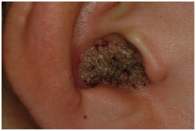
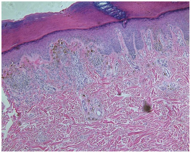
A 47-year-old Korean man presented with a 1-year history of a well-demarcated 1├Ś1 cm, dark-brown to black papillomatous plaque in his right cavum conchae (Fig. 1). He didn't complain of any pain, itching, intermittent oozing, or hearing disturbance. On otoscopic examination, his right tympanic membrane and external auditory canal were normal. He was healthy and had no history of skin cancer or other skin problem. A skin biopsy specimen showed numerous nests and clumps of melanocytic nevus cells in the upper dermis and dermoepidermal junction (Fig. 2). The lesion was treated with CO2 laser (Model 1020C 20WA, Sharplan Lasers Inc., Migdal Haemek, Israel) abrasion at 1.0 watt in the super-pulsed mode. Only topical antibiotic ointment without coverage was applied for post-laser wound care for 1 week. The patient showed no recurrence during the 6-month follow-up period (Fig. 3).
Discussion
The common acquired melanocytic nevus is a benign neoplastic proliferation of melanocytes. Histologically it is diagnosed by the presence of nevus cells that are arranged in clusters or nests, and is classified into junctional, compound, and intradermal nevi, in which the nevus cells are confined to the epidermis, epidermis and dermis, and dermis respectively. In addition to the histological classification, five clinical types of lesion can be recognized: 1) flat, 2) slightly elevated often with raised centers and flat peripheries, 3) papillomatous, 4) dome-shaped, and 5) pedunculated.1) In most cases, slightly elevated lesions represent compound nevi, while papillomatous, dome-shaped, and pedunculated lesions represent intradermal nevi. Friedmann2) reported the first case of melanocytic nevus in the skin of the external auditory canal and two cases of melanocytic nevi presenting as papillomatous lesions that were revealed as intradermal nevi have been described in the English literature.3,4) In our case, the lesion was a papillomatous plaque similar to the previously reported cases, but it turned out to be a compound nevus.
Melanocytic nevi occurring in the auricular region, like those occurring in the acral, flexural, or genital area, may exhibit some of the histologic features commonly found in melanoma such as pagetoid spread of melanocytes, cytologic atypia, and asymmetric growth pattern.5) Therefore, careful histologic interpretation of these rare lesions is recommended. The differential diagnosis should include viral wart, seborrheic keratosis, foreign body granuloma, and a variety of benign and malignant neoplasms.
The CO2 laser has been widely used for the removal of various skin tumors, including melanocytic nevus. However, it was usually effective only in small superficially located tumors with surgical excision being recommended for large or deeply seated tumors. As mentioned above, the wide surgical excision of nevi occurring near the external auditory canal is difficult, and hence we attempted CO2 laser abrasion despite the large size in the case presented here. We had expected several treatments to be necessary, but found that a single session of treatment was sufficient for clearance.
To the best of our knowledge, this is the first case of a compound papillomatous nevus occurring near the external ear canal. We consider that compound nevus should be included in the differential diagnosis of papillomatous skin lesion occurring near the external auditory canal and that CO2 laser abrasion can be a good choice for its treatment.